Because of the origin of the retinae and optic nerves from the developing forebrain, the optic nerves (cranial nerves II) corresponds to fibre tracts connecting parts of the CNS - in this case the ganglion cells of the retina with neurones in the lateral geniculate nucleus of the thalamus and neurones in the superior colliculus and pretectum of the midbrain.
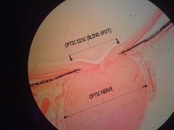
Ganglions cell axons run towards the optic disc where they turn towards the sclera. Numerous bundles (or fascicles) of axons pass through the choroid and openings in the sclera, thelamina cribosa. The axons become myelinated in this region. Collectively, the bundles form the optic nerve. Like other parts of the CNS, the optic nerve is surrounded by the three meninges - the outer dura mater, the middle archnoid and the inner pia mater, which are separated from each other by subdural and subarachnoid spaces. At the eyeball, the dura fuses with the sclera while the arachnoid and pia mater merge with the choroid. Connective tissue septa, which arise from the pia mater, separate the fibre bundles in the optic nerve. The axons in the optic nerve are supported by astrocytes and oligodendrocytes. Microglia is also present.
Reference: www.lab.anhb.uwa.edu.au